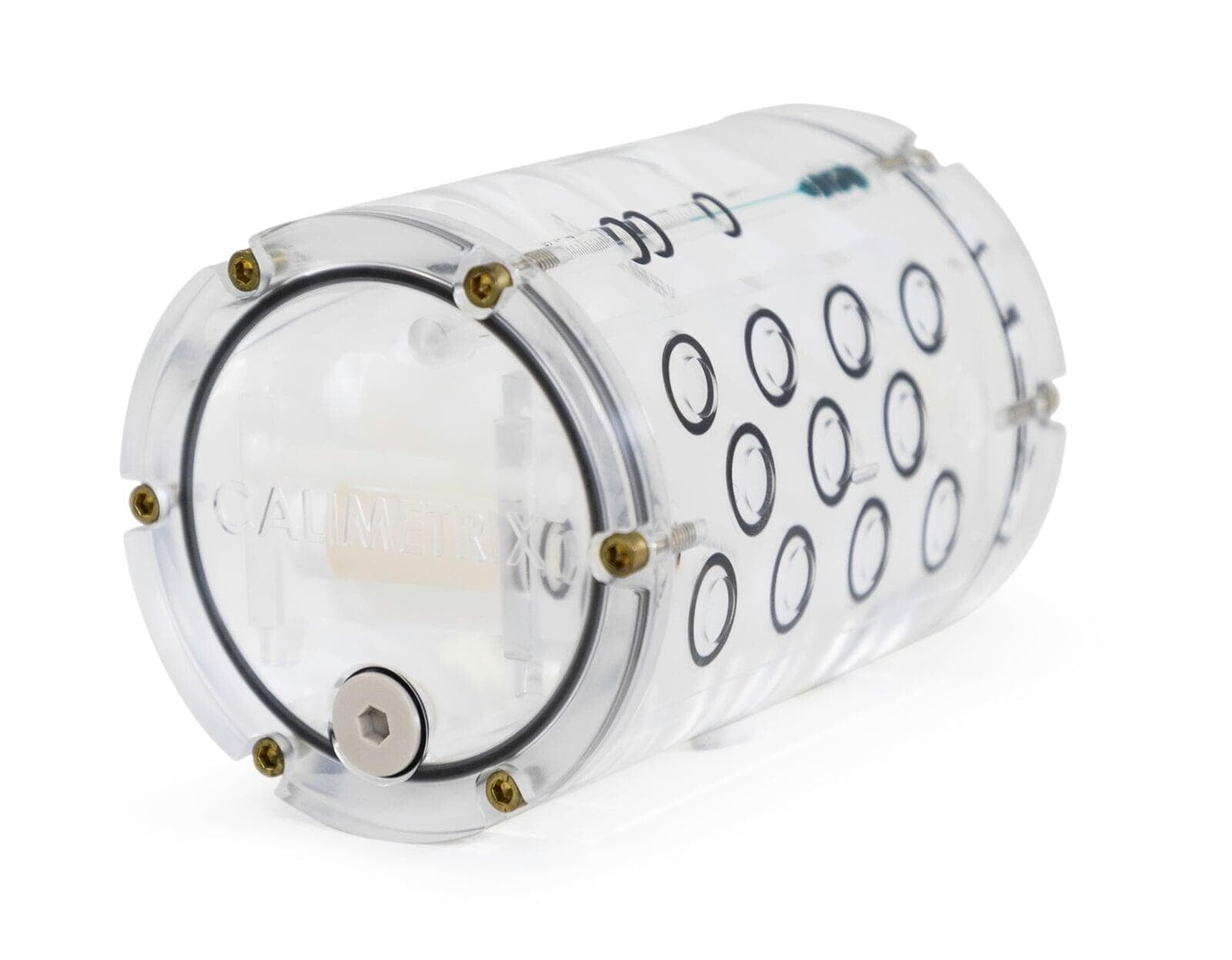
MSK Phantom Features
- Contains 12 gel-based vials
- Nominal (target) T1/T2 values when measured at 3T: 1200/10, 1200/20, 1200/30, 1200/40, 1200/50, 1200/60, 1200/70, 1200/80, 1200/90, 1200/100, 700/40, 300/40 ms.
- Asymmetrically positioned vials to avoid left-right and superior-inferior vial identification ambiguities
- Doped fill solution to improve B0 and B1 homogeneity
- The long axis of the vials is oriented perpendicular to the axis of the housing to enable the sagittal and coronal slice orientation typically used in knee imaging
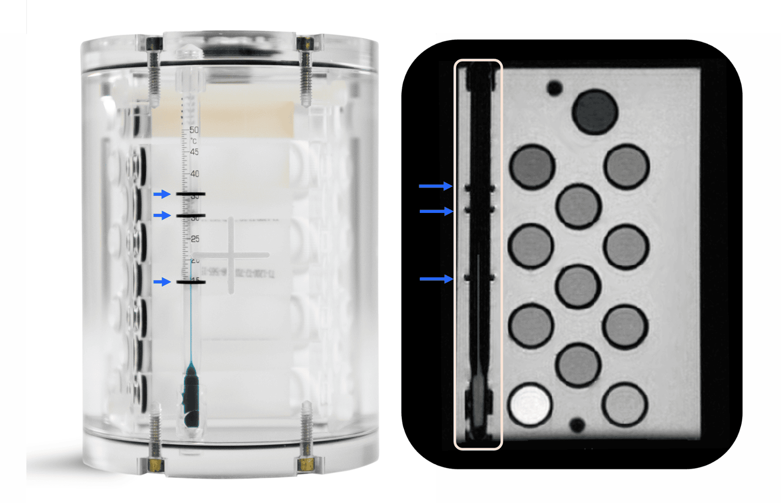
Embedded Temperature Reference
The phantom contains a thermometer that can be utilized by (1) physically observing the thermometer through the clear phantom wall before or after imaging and/or (2) reading the thermometer on the phantom MR images. The phantom temperature can be estimated from the phantom images by noting the position of the thermometer liquid column relative to the three dark bands created on the thermometer by the thermometer markers
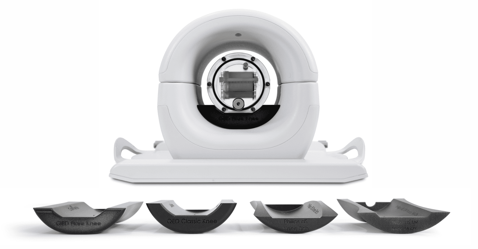
Custom Stands for Common Knee Coils
Knee coils often have curved internal surfaces, making consistent phantom placement difficult. Therefore, we offer phantom stands designed to approximately center the phantom in the following MRI coils:
- Philips "dS T/R 16ch" knee coil
- Philips "dS Knee 8ch" knee coil
- QED knee coils with "QED Classic" design
- QED knee coils with "QED Flare" design
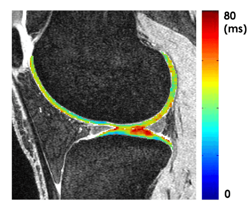
Clinical Relevance of Musculoskeletal Relaxometry
Clinical Relevance of Musculoskeletal Relaxometry
Quantitative musculoskeletal relaxometry imaging is used to assess tissue compositional changes at an earlier stage than they can be detected with traditional qualitative imaging. For example, by measuring parameters such as T1, T2, and T1rho, early cartilage degeneration in osteoarthritis can be identified. Such quantitative relaxometry measurements can be sensitive to imaging parameters and system changes over time. Therefore, the Calimetrix MSK Relaxometry Phantom was developed to provide a quality assurance tool that can be used to standardize and track MRI relaxometry measurements over time and between systems and methods.