6 Vials: 0, 10, 20, 30, 40, 100% PDFF
12 Vials: 0, 2.5, 5.0, 7.5, 10, 15, 20, 25, 30, 40, 50, and 100% PDFF
18 Vials: 0, 2.5, 5, 7.5, 10, 15, 20, 30, 40, 50, 60, 70, 75, 80, 85, 90, 95,100% PDFF
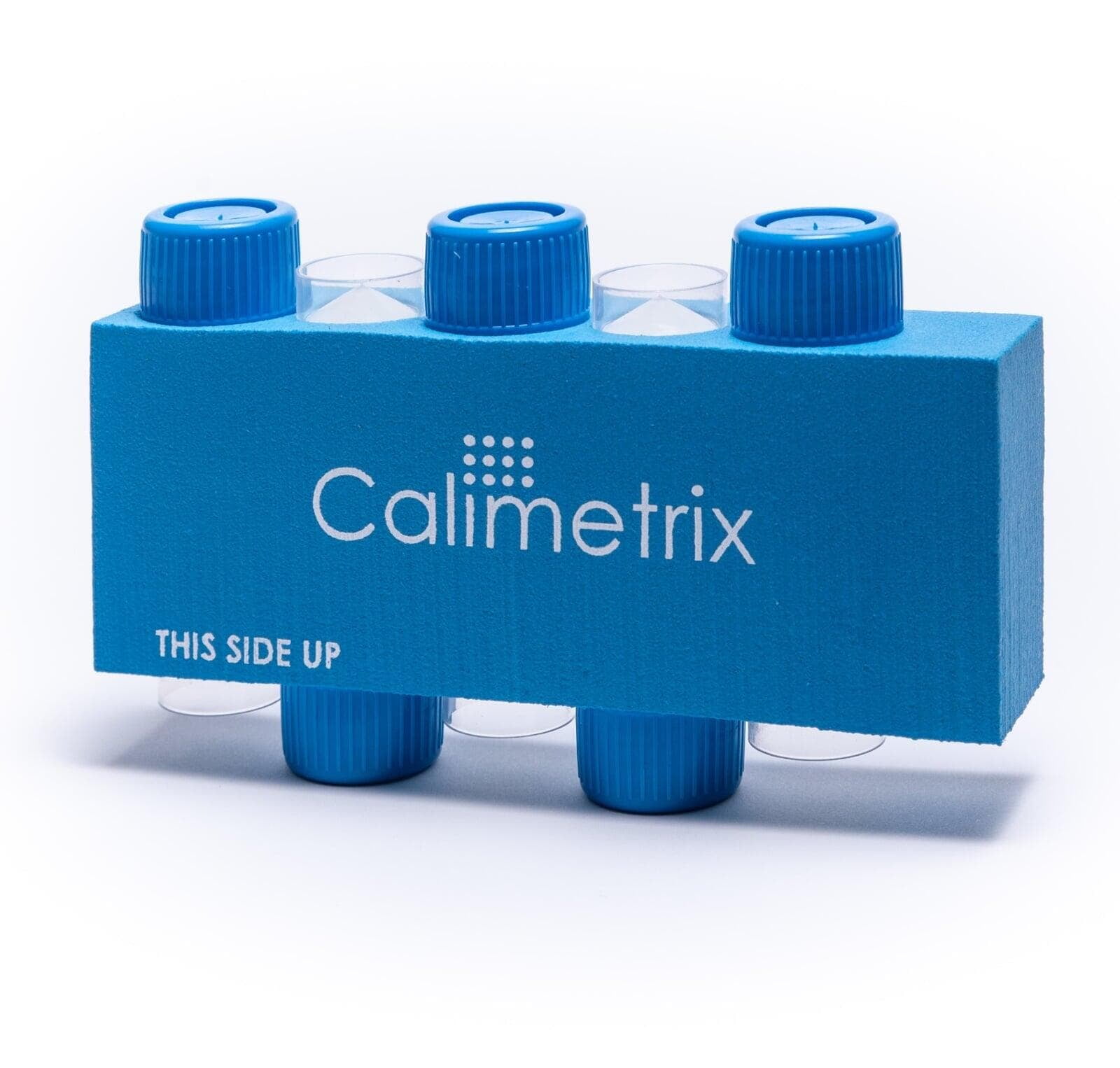
5 Vials: 0, 10, 20, 30, 40% PDFF. Shown with optional accessory pad
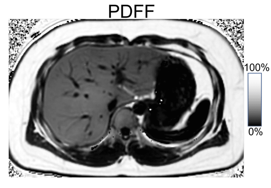
Clinical Relevance of PDFF Quantification
MRI-based triglyceride metrics can be useful for diagnosis, quantitative grading, and treatment monitoring of metabolic dysfunction–associated steatotic liver disease (MASLD), previously known as non-alcoholic fatty liver disease (NAFLD), and other diseases. However, such quantitative metrics must be both accurate and reproducible. Our Model 300, Model 600, and Phantom Pack products focus on the 0-50% PDFF range that is relevant for MASLD. Our Model 825 product includes PDFF values that sample the complete range from 0-100% PDFF. This is relevant for the following tissues:
- Fatty Liver Disease: 0-50% PDFF
- Fatty Infiltration of Muscle: 0-15% PDFF
- White Adipose Tissue (e.g. Subcutaneous Fat): 75-100% PDFF
- Brown Adipose Tissue: 20-85% PDFF
- Red Bone Marrow: 20-70% PDFF
Relevant Publications
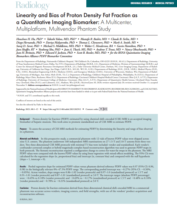
Hu, H.H., Chen, H.SM. & Hernando, D. Linearity and bias of proton density fat fraction across the full dynamic range of 0–100%: a multiplatform, multivendor phantom study using 1.5T and 3T MRI at two sites. Magn Reson Mater Phy (2024). https://doi.org/10.1007/s10334-024-01148-9
Stelter, J.K., Boehm, C., Ruschke, S., Weiss, K., Diefenbach, M.N., Wu, M., Borde, T., Schmidt, G.P., Makowski, M.R., Fallenberg, E.M., Karampinos, D.C., Hierarchical multi-resolution graph-cuts for water–fat–silicone separation in breast MRI. IEEE Transactions on Medical Imaging 11, 3253–3265. (2022)
Zhao R, Hamilton G, Brittain JH, Reeder SB, Hernando D. Design and evaluation of quantitative MRI phantoms to mimic the simultaneous presence of fat, iron, and fibrosis in the liver. Magn Reson Med. 2021 Feb;85(2):734-747. doi: 10.1002/mrm.28452. Epub 2020 Aug 12. PMID: 32783200
Tang A, Desai A, Hamilton G, Wolfson T, Gamst A, Lam J, Clark L, Hooker J, Chavez T, Ang BD, Middleton MS, Peterson M, Loomba R, Sirlin CB. Accuracy of MR imaging-estimated proton density fat fraction for classification of dichotomized histologic steatosis grades in nonalcoholic fatty liver disease. Radiology. 2015 Feb;274(2):416-25. doi: 10.1148/radiol.14140754. Epub 2014 Sep 22. PMID: 25247408; PMCID: PMC4314291.
Reeder SB, Hu HH, Sirlin CB. Proton density fat-fraction: a standardized MR-based biomarker of tissue fat concentration. J Magn Reson Imaging. 2012 Nov;36(5):1011-4. doi: 10.1002/jmri.23741. Epub 2012 Jul 6. PMID: 22777847; PMCID: PMC4779595.
Reeder SB, Hines CD, Yu H, McKenzie CS, Brittain JH. On the Definition of Fat-Fraction for In Vivo Fat Quantification with Magnetic Resonance Imaging. Proc. Intl. Soc. Mag. Reson. Med. 17 (2009).