PDFF-R2* Phantom (Model 725)

The Calimetrix PDFF-R2* Phantom simultaneously controls PDFF and R2* in each vial. It is designed for testing and quality control of magnetic resonance (MR)-based fat and iron quantification.

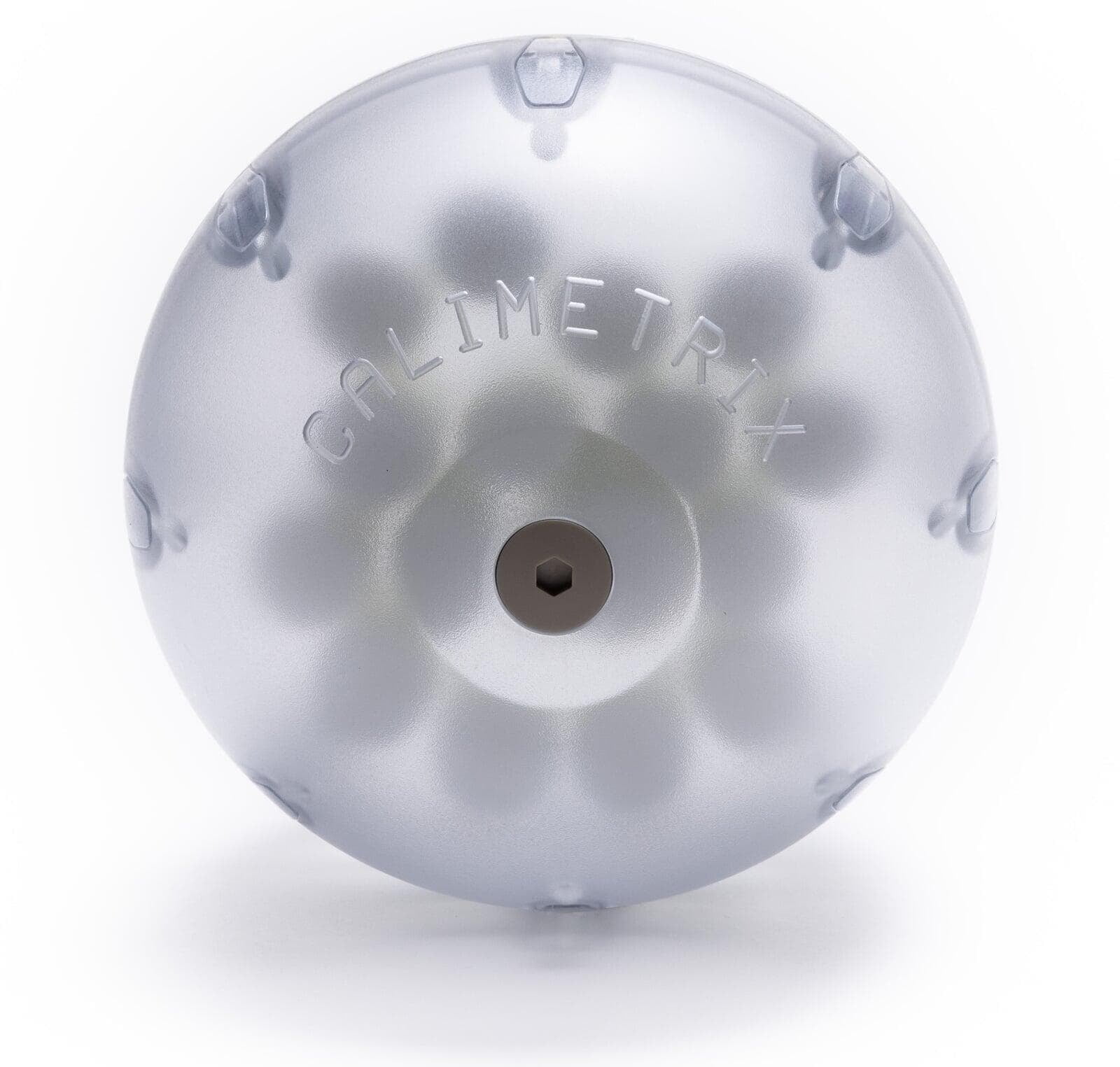
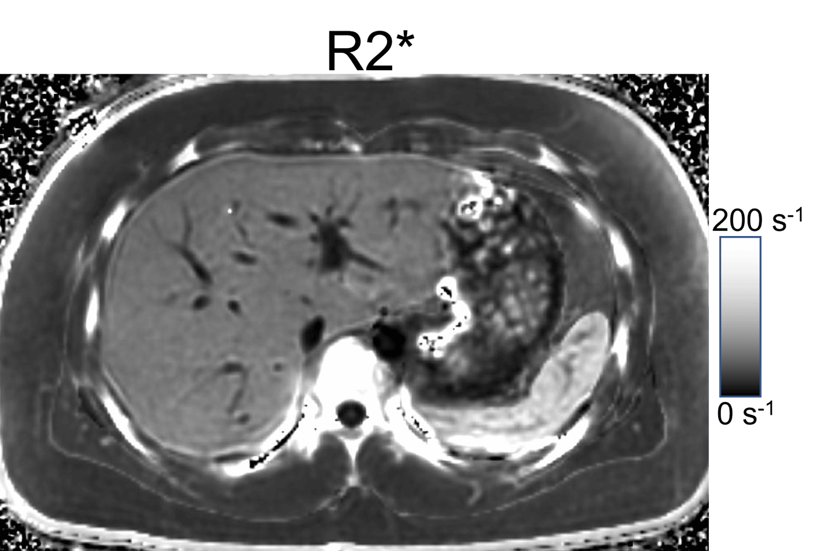
Model 725 PDFF-R2* Phantom Features

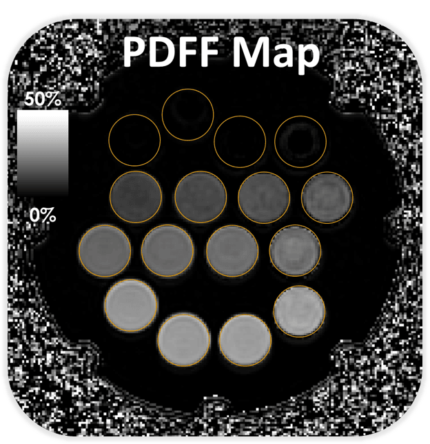
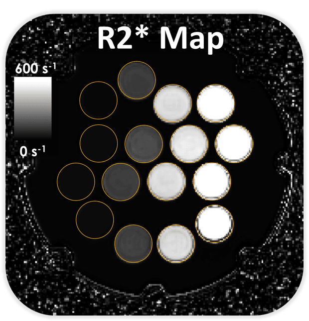
  • 16 Vials: (0/50), (0/150), (0/350), (0,600), (10/50), (10/150), (10/350), (10,600), (20/50), (20/150), (20/350), (20,600), (30/50), (30/150), (30/350), (30,600) [%PDFF/s-1] at 3T

  • Phantom vials are filled with gels to minimize motion-related artifacts

  • Asymmetrically positioned vials to avoid left-right and superior-inferior vial identification ambiguities

  • Doped fill solution to minimize water-fat swap artifacts

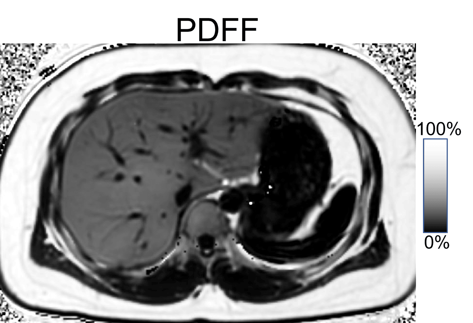
Clinical Relevance of PDFF-R2* Quantification

Abnormal fat and iron deposition in the liver are principal histological features of many diseases. For example, non-alcoholic fatty liver disease (NAFLD), also known as steatotic liver disease (SLD), is emerging as the leading cause of liver disease. Iron overload often coexists with NAFLD, and liver iron is also an important indicator of total body iron in patients with genetic hemochromatosis and those who receive multiple blood transfusions. Abnormal liver fat and iron overload lead to liver injury and fibrosis, and eventually cirrhosis, liver failure and hepatocellular carcinoma. Importantly, fat and iron also commonly co-exist as key histological features of many other diseases involving the heart, pancreas, kidneys, among others. In recent years, noninvasive MRI biomarkers of fat (based on proton density fat fraction, ie: PDFF) and iron (based on R2*) have become available. Chemical shift encoded MRI methods estimate PDFF and R2* from the same multi-echo data set.  This is important because the R2* signal decay rate, which increases linearly with iron concentration, is well known to confound the quantification of fat, if not accounted for in the signal estimation models. Similarly, the presence of fat is well known to confound the ability to quantify iron.  Our PDFF-R2* phantom enables more accurate testing and quality assurance of methods to quantify PDFF and R2*.

Relevant Publications

Zhao R, Hamilton G, Brittain JH, Reeder SB, Hernando D. Design and evaluation of quantitative MRI phantoms to mimic the simultaneous presence of fat, iron, and fibrosis in the liver. Magn Reson Med. 2021 Feb;85(2):734-747. doi: 10.1002/mrm.28452. Epub 2020 Aug 12. PMID: 32783200

Starekova J, Rutkowski D, Bae W, Do H, Madhuranthakam A, Malis V, Mukherjee S, Qin Lin S, Serai S, Yokoo T, Reeder S, Brittain J, Hernando D. Muli-center, multi-vendor validation of PDFF-R2* mapping in an optimized fat-iron phantom. International society for magnetic resonance in medicine. 2023. Toronto, ON, Canada. Summa Cum Laude Merit Award.