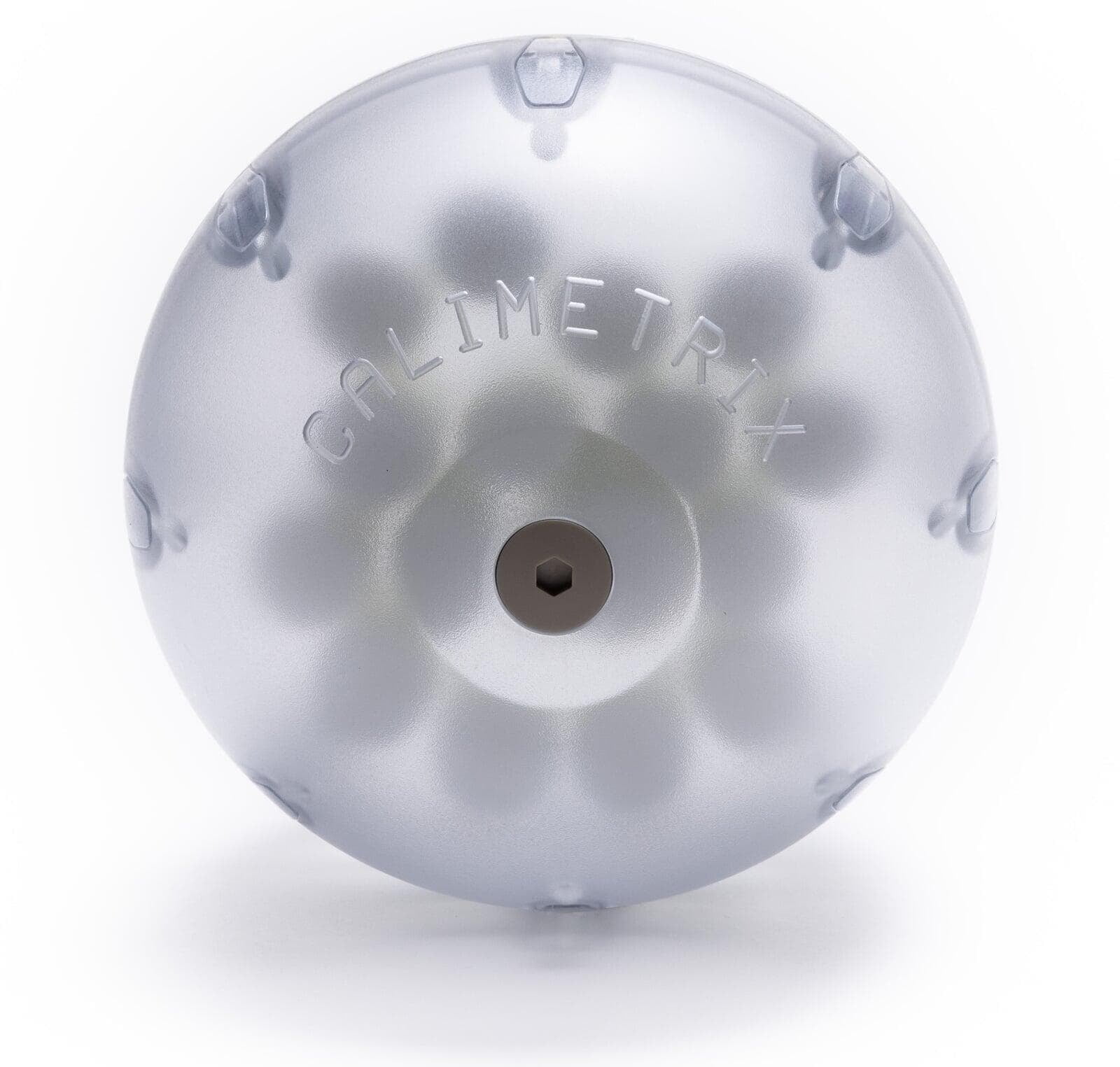
Special Order PDFF-T1 Phantom Features
- 16 Vials: (0/200), (0/600), (0/1000), (0/1400), (10/200), (10/600), (10/1000), (10/1400), (20/200), (20/600), (20/1000), (20/1400), (30/200), (30/600), (30/1000), (30/1400) [%PDFF/ms] at 3T
- Phantom vials are filled with gels to minimize motion-related artifacts
- Asymmetrically positioned vials to avoid left-right and superior-inferior vial identification ambiguities
- Doped fill solution to minimize water-fat swap artifacts
Clinical Relevance of PDFF-T1 Quantification
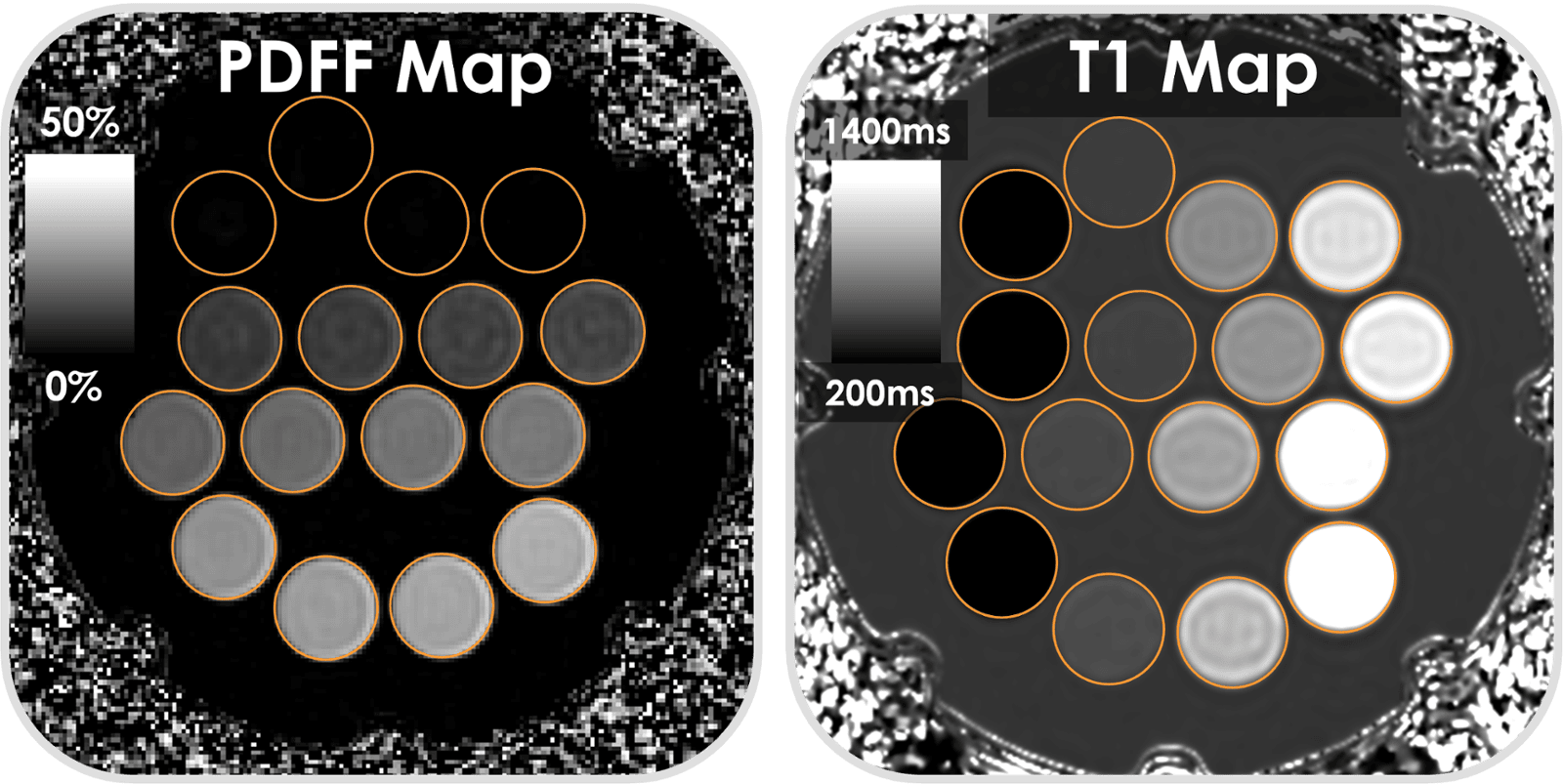
MRI-based PDFF mapping is widely accepted as the most accurate and precise noninvasive biomarker for the evaluation of liver steatosis. In addition, T1 mapping has emerged as a promising noninvasive biomarker for liver fibrosis and inflammation (native T1) and liver function (postcontrast T1 using hepatocyte-specific contrast agents). Importantly, the presence of steatosis can confound T1 mapping, and the presence of T1-related bias can confound PDFF mapping. The Calimetrix Special Order PDFF-T1 Phantom enables quality assurance of these important biomarkers.
Relevant Publications
Starekova J, Weingärtner S, Rutkowski DR, et al., “Cross-Vendor Validation of Proton Density Fat Fraction and T1 Mapping Using a Combined Proton Density Fat Fraction—T1 Phantom,” Journal of Magnetic Resonance Imaging (2026): 1–11, https://doi.org/10.1002/jmri.70344.
Zhao R, Hamilton G, Brittain JH, Reeder SB, Hernando D. Design and evaluation of quantitative MRI phantoms to mimic the simultaneous presence of fat, iron, and fibrosis in the liver. Magn Reson Med. 2021 Feb;85(2):734-747. doi: 10.1002/mrm.28452. Epub 2020 Aug 12. PMID: 32783200
Starekova J, Weingärtner S, Rutkowski D, et al. Multi-center, multi-vendor validation of PDFF and T1 mapping in an optimized PDFF-T1 phantom. International Society for Magnetic Resonance in Medicine Annual Meeting. May 15-20, 2021 2024;